
Unusual Sites of a Common Pathogen: Giardiasis Diagnosed on Terminal Ileum and Colon Biopsies
By Reenu Malhotra, M.D.
Trophozoites of Giardia lamblia can rarely be found on microscopic examination of colon biopsies which are performed for unrelated reasons. In our practice we recently encountered three patients who underwent colonoscopy for varied gastrointestinal symptoms and Giardia trophozoites were identified on colon and terminal ileum biopsies.
Of these three patients, one young male patient (23 yrs) came in with history of diarrhea, one (40 yrs. male) had hematochezia and one patient (60 yrs. male) came in for screening. These three
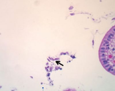
Fig. 1: Terminal ileal mucosa with Giardia trophozoites
patients underwent colonoscopy with biopsies of colonic mucosa, and one underwent terminal ileal biopsy as well. All the biopsies revealed presence of pear and comma shaped proto-zoal organisms consistent with Giardia Lamblia. The colonic mucosa in two of the patients was otherwise unremarkable with normal plasma cell population, no increased intraepithelial lymphocytes and no active or chronic inflammatory injury. The 23-year male who showed presence of Giardia lamblia in colonic and terminal ileal biopsy (Fig. 1) showed patchy increase in surface intraepithelial lymphocytes at both places (Fig 2). There was no duodenal biopsy done in any of these patients, as possibility of Giardia infection was not considered clinically.
Worldwide, Giardia is the most common protozoan isolated from the gastrointestinal tract (1). Orally trans-mitted as a cyst, it usually infects the proximal small bowel, the common site for trophozoites, because excystation is aided by gastric acid (1-3). Giardia lamblia intestinal infection is not usually associated with mucosal inflammation or lesions. Mild villous short-ening and mild non‐specific inflammation of the lamina propria have been reported in less than 5% of patients with duodenal infection (1). Duodenal colonization may be responsible for acute and chronic malabsorption and secretory diarrhea is the suggested pathogenetic mechanism of the disease. Encystation of G lamblia trophozoites occurs in the small bowel. Cysts are then excreted with stools. It is therefore not common to see trophozoites in the colon (1) or in stools (2).
Colonic giardiasis has been rarely reported in immunocompetent hosts (1,2). In a series of 567 patients diagnosed with G lamblia, the site of colonization was the small bowel in most cases (97%), but rarely the colon (two patients (0.4%). In another series of 124 patients diagnosed with G lamblia positive stools, 120 had only cysts (97%), four had trophozoites and cysts (3%) and none had trophozoites only
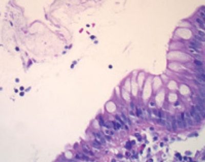
Fig. 2: Increased intraepithelial lymphocytes with Giardia trophozoites (Colon Biopsy)
(2).
The contributing factors are speculative. Cholesterol deprivation and exposure to bile acids are demonstrated in vitro as two major stimuli of G lamblia encystation (3). Similarly, in vivo, cholestasis and diet supplemented with cholestyramine have also been shown to dramatically decrease the fecal excretion of G muris cysts in mice (4). The findings suggest that fibrate treatment, which increases biliary cholesterol excretion and decreases the excretion of bile acids, hampered giardia encystations in these patients, subsequently facilitating G lamblia colonic colonization of trophozoites. In addition, changes in bile metabolism induced by hypolipidemic drugs (not only fibrates but also niacin and statins) or bile acid resins (cholestyramine) may influence the presentation and spread of G lamblia (5).
Regardless of whether these cases represent true colonic infection, it is of interest that Giardiasis was diag-nosed from ileal and colonic biopsies (Fig. 1, 2 & 3). Pathologists should be aware of this rare finding at these unusual locations as correct diagnosis can be rendered and a simple treatment can be instituted.
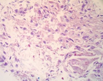
Fig. 3: Trophozoites on luminal surface of colon biopsy
References
1. Oberhuber G, Kastner N, Stolte M. Giardiasis: a histologic analysis of 567 cases. Scand J Gastroenterol 19973248–51.
2. Howard L H, Fink D S, Lubin J.et al Giardiasis diagnosed by biopsy of the colon and terminal ileum: unusual sites for a common pathogen. Am J Gastroenterol 1995901011–1013.
3. Lujan H D, Mowatt M R, Byrd L G.et al Cholesterol starvation induces differentiation of the intestinal parasite Giardia lamblia. Proc Natl Acad Sci USA 1996937628–7633.
4. Erlandsen S. Reduction in fecal excretion of Giardia cysts: effect of cholestasis and diet. J Parasitol 20059 11482–1484.
5. X Dray, V Medeau, M Qubaja et al: Colonic colonization with Giardia lamblia in a patient receiving fibrates. Gut. 2007 Nov; 56(11): 1639–1640.